
Manuela Callari
We are taking a look back at stories from Cosmos Magazine in print. In March 2024, Manuela Callari reported on the burgeoning excitement around bionic eye technology and the doubts that linger about its ability to truly restore sight.
When Colleen Knowles heard that researchers at the Royal Victorian Eye and Ear Hospital, in Melbourne, were looking for volunteers to trial a bionic eye prototype, she signed up without hesitation. “I have always liked history – I thought I could be part of it,” she says.
Knowles was born short-sighted. She saw through thick glasses from a young age, but her sight deteriorated and her world went dark by age 30. In 2017, when she was 66, doctors placed a bionic implant – also known as a visual prosthesis – in her right eye; her sight wasn’t fully restored, not by a long shot, but rough shapes and patterns of lights replaced the darkness.
“I like going to a shopping plaza and watching people go by and try to work out what shops they’re going into,” Knowles says.
The excitement around eye implants has grown over the past two decades, with more than 40 research teams worldwide working on bionic vision. The global bionic eye market was valued at more than US$297 million (about $427 million) in 2022 and is expected to reach US$558.36 million (about $860 million) by 2028.
Several eye implants have been tested in people. None have succeeded in significantly restoring vision, making some wonder if the excitement about this field of research is still justified. How close are we to a bionic eye that genuinely restores sight? Ironically, given the rapid advances in genetic therapies, bionic eyes might become outdated gadgets before they even hit the market.
Losing sight
When light hits Colleen Knowles’ eyes, it triggers no response in her retinas, so no signals flash up her optic nerves to her brain. A rare genetic disease called cone-rod dystrophy killed the photoreceptor cells in her retinas.
In Australia, around 16,000 people are living with an inherited retinal disease. Cone-rod dystrophy primarily affects cones, followed by rods. It starts with a loss of central vision and colour perception, leading to difficulties with tasks like reading or recognising faces. But in retinitis pigmentosa – the most common inherited retinal disease – the initial damage occurs in rods, followed by cones. It often begins with night blindness, followed by tunnel vision as the peripheral vision deteriorates. It affects an estimated one in every 5,000 people worldwide, and one in every 3,000 people in Australia, which equates to about 8,500 Australians.
These conditions are caused by genetic anomalies people have at birth that lead to the degeneration of the rods and cones. The faulty gene produces a dysfunctional protein (or no necessary protein) for maintaining the function and structure of photoreceptor cells in the retina.
There’s no known cure for these diseases. But technology might come to the rescue.
While these conditions damage the rods and cones, the remaining retinal elements within the inner retinal layers survive in large numbers and remain responsive to electrical stimulation even in highly advanced stages of the disease. These elements include the bipolar and ganglion cells. Bipolar cells transmit signals from the photoreceptor rods and cones to the ganglion cells, which in turn pass that information along to several regions of the brain.
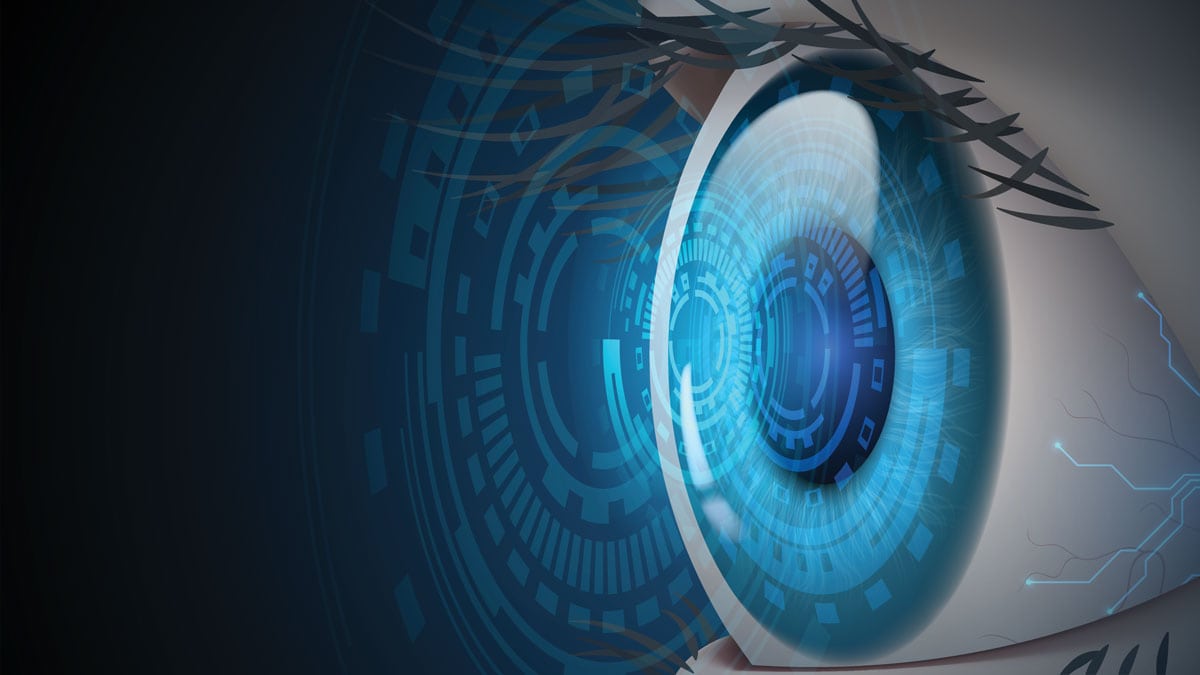
The aim of bionic implants is to replace the function of the degenerated photoreceptor cells by artificially stimulating the surviving retinal neuronal machinery.

Restoring sight
Most eye implants have the same backbone, with only subtle differences in the type of surgery involved in placing them.
The typical model employs an external camera mounted on a pair of spectacles to capture visual information from the environment. The captured images are processed by a small unit, often worn by the user, which converts the images into signals that the brain can understand. The signals are transmitted wirelessly to a receiver placed under the patient’s skin next to the eye and wired to an array of electrodes implanted in the eye either in front of or behind the retina. The electrodes bypass the damaged photoreceptor cells and stimulate the remaining healthy nerve cells, triggering them to send signals to the brain.
Perhaps not surprisingly, interest in developing visual prostheses grew from the transformational success of cochlear implants. In the late 1990s, biomedical engineer Robert Greenberg founded California-based Second Sight Medical Products. Among Greenberg’s investors was a multimillionaire named Sam Williams, who was blind due to retinitis pigmentosa.
In a FDA-mandated post-approval study, Second Sight reported 36 severe and 152 non-serious adverse events.
After extensive testing and animal trials, Second Sight’s first implant device, called the Argus I, was placed in the eyes of a 74-year-old US volunteer in 2002. Another five volunteer testers followed over the next three years. The Argus I was a basic 16-electrode device that allowed patients to see if the room was dark or light or if someone was moving in front of them as well as recognise some simple shapes on a screen.
Second Sight’s next-generation, 60-electrode Argus II rolled out in 2006; 30 patients around the world were implanted with the device over the next few years. But what they could see didn’t improve by much.
In 2011, the Argus II was approved for commercial use in the European Union, and two years later was authorised by the US Food and Drug Administration (FDA) – under a humanitarian device exemption – for use by up to 4,000 people. But by 2019, Second Sight had discontinued its retinal implant and gone out of business, leaving the more than 350 blind people around the world implanted with the Argus device in the dark. Without assistance or spare parts available, one broken wire means a patient will be left with a defunct device in their eye forever.
Second Sight merged with another medical tech company now called Vivani Medical. Uday Patel, Director of Clinical and Scientific Affairs at Cortigen, a subsidiary of Vivani, says that at $150,000, the device was too expensive and the market was too small to make a profitable business. He says phasing out the project was a business decision despite successful results. However, in a post-approval study mandated by the FDA that followed 30 patients from 2007 to 2019, Second Sight reported 36 severe and 152 non-serious adverse events.

“The Argus did not work well,” says Ash Attia, CEO at Bionic Vision Technologies (BVT), a commercial spinoff from the Bionic Vision Australia (BVA) research consortium.
The problem, according to Attia, lies in the position of the array of electrodes and the surgical procedure to place it. With epi-retinal implants like the Argus, the surgeon must detach the retina and stick the array of electrodes in front of it, destroying residual photoreceptors. “Anytime you stick something on in the body, you cause damage,” Attia says.
Researchers at BVT have developed a suprachoroidal electrode array that is inserted in a natural cleavage plane between the choroid and the sclera. The choroid provides oxygen and other nourishment to the retina; it’s also rich in the dark pigment melanin, which limits uncontrolled reflection in the eye that might create the perception of confusing images. The sclera is the eye’s outer protective layer, which mainly contains collagen.
To insert the suprachoroidal electrode array, the surgeon opens the wall of the eye, dissects the suprachoroidal space and slides in the array without touching the retina. “We go into a biological space,” says Attia. “We are not sticking anything on cells. It is a very simple surgery that preserves any remaining photoreceptors.”
A risk of going into the suprachoroidal space is causing haemorrhage bleeding – because it is rich in blood vessels that carry oxygen and nutrients to the normally highly active photoreceptor cells. “That is not the case for these patients,” says Penelope Allen, an associate professor at the University of Melbourne and Principal Investigator at the Centre for Eye Research Australia (CERA). “Their photoreceptors have died, and the blood vessel layer is actually quite atrophic.”
The first proof-of-concept study conducted by BVA began in March 2012. Researchers implanted a 24-electrode array into the suprachoroidal space behind the retina of three patients with only light perception vision. This first trial looked at whether the implant could generate visual perception in people with profound vision loss. Patients could only use the device one day a week at the Royal Victoria Eye and Ear Hospital, and at the end of the two-year study, they had the external wires connected to the implant removed.
How our nerves send signals is complicated, and making devices that understand this complexity is tough.
This initial success saw BVT move on to a second trial. In 2017, they implanted Knowles and three other patients with retinitis pigmentosa with their second-generation device. The new 44-electrode bionic eye was fully implantable, which meant patients could go home with it. “We have had excellent, excellent results,” says Attia.
“Four years post-implantation, the devices are stable in position, and we have had no ocular complications,” says Allen. “Obviously, it’s a small group, but results are certainly very positive.”

The Melbourne-based start-up received $1 million from the Medical Research Future Fund’s BioMedTech Horizons 3.0 program in late 2020. The funds support the development of vision-processing software to add features like depth perception.
The new software is a key part of BVT’s next-generation device, which is set to be used in a global study to gather data to support regulatory submissions in major commercial markets.
“We are gearing up for the very last trial,” says Attia. “Once [the device] gets regulatory approval, we’ll look into reimbursement, because those patients can become independent again.”

Not quite the vision we’d hoped for
Six weeks after surgery, it was “switch-on day” for Colleen Knowles. She sat patiently as engineers and doctors worked around her head to connect her device to a computer. Once everything was set up, the research crew sent electrical stimulation to Knowles’ device, hoping to reignite the dormant pathways of her sight.
“At first, I didn’t think it was going to work,” recalls Knowles. But as electrical stimulation increased, flickers of light emerged in her long-darkened world. “It was quite exciting.”
It took Knowles two months to make sense of these flashes. “These patients have had no useful vision for over 15 or 20 years,” says Allen. “They need to learn to interpret the stimulation.”
Eventually, Knowles learnt to get through obstacle courses. She could identify if a door was open or shut or if someone she was talking to had walked away. “It’s not to the point where I can replace my guide dog,” she says. “But I can tell if I’m looking at a tree or an electricity pole.”
While the device gives Knowles a perception of her surroundings, it does not give her the experience of colours, depth and movements.
“Vision is a very complex sense, and these devices are too basic,” says Gregg Suaning, a biomedical engineer at the University of Sydney.
Suaning says one of the most difficult challenges is figuring out how the brain interprets what we see. “How our nerves send signals is complicated, and making devices that understand this complexity is tough.”
We need to be honest about what these devices can and cannot deliver … It’s not as exciting as we thought.
In comparison, when paired with cochlear implants, the brain compensates for missing auditory data. “Your brain is amazing at adapting to sounds,” Suaning says. “But, for reasons I don’t fully understand, the brain isn’t really good at interpreting a whole lot of new things with vision.”
There are two major limitations to the resolution that eye implants can provide. The first is the distance between the electrodes and the nerve cells – the further apart they are, the higher the electrical stimulation must be. The second is the relationship between electrode size and electrical current output. A certain current is required to stimulate the nerve cells, and smaller electrodes deliver less of it, while large electrodes tend to activate multiple neurones at the same time, which may send conflicting signals that the brain struggles to interpret. To achieve higher resolution, the device’s electrodes must be both smaller and capable of delivering more current.
University of Sydney researchers, led by Suaning, have developed a device called Phoenix99, where the electrodes are little pillars of about 200 micrometres that penetrate the nerve cell layer from behind the retina. “That, theoretically, will make the stimulation more circular and small so that we could pack more electrodes into a small area and deliver a lot more meaningful vision,” says Suaning.
Novel therapies advancement
While some researchers figure out the best way to stimulate optic nerve cells, others are developing ways to fix or suppress the faulty genes that cause diseases like retinitis pigmentosa. Such gene therapies have come a long way in recent years.
Scientists use viruses, such as adeno-associated viruses (AAV), to deliver functional genetic material to retinal cells, helping them produce the missing or defective proteins again. AAVs are a top choice because they are small, less likely to cause immune reactions and can effectively target the retina. In 2017, Luxturna (voretigene neparvovec-rzyl) was the first vision gene therapy approved by the FDA. Clinical trials have shown that patients receiving Luxturna exhibit signs of vision improvement, such as enhanced light sensitivity and better visual acuity. Developed by Spark Therapeutics, it costs a jaw-dropping $850,000 for both eyes, which may be covered by insurance.
However, Luxturna is only suitable for patients with a mutation in both copies of the RPE65 gene, representing 0.3–1% of all retinitis pigmentosa cases. A single injection of Luxturna delivers a healthy copy of RPE65 directly to the retina, restoring its ability to respond to light.

Many companies are targeting other genes responsible for different kinds of retinitis pigmentosa, with promising results in halting progressive vision loss and, in some cases, improving visual capabilities. Biotech firm MeiraGTx, for example, is developing a gene therapy for treating patients with X-linked retinitis pigmentosa (XLRP) with disease-causing variants in the RPGR gene. Interest in gene-editing technologies to cure diseases is growing. But scientific and biological challenges, as well as the enormous costs of some of these therapies, persist, says Anna Greka, Director of the Kidney Disease Initiative at the Broad Institute of MIT and Harvard.
Greka and her team are looking at pills to treat genetic diseases such as retinitis pigmentosa.
In 1960, surgeon Roscoe Nelson died from a mysterious disease. He knew his kidneys were progressively failing but had no idea of the cause, and no access to treatment. Several years later, four of his six children also developed kidney failure. Now that scientists are able to read every letter in the human genome, Greka and her team could identify a mutation in a gene called MUC1 in the Nelsons’ DNA. The mutation causes the production of a misshapen protein that binds to another molecule called TMED9 and accumulates inside kidney cells. “Over time, this becomes toxic to the kidney cells, and the cells die,” Greka explains.

Her team was excited when they found a drug that – in mice – can direct this toxic waste into the lysosomes, the cell’s trash disposal system. But they also found “that the exact same entrapment mechanism is true [for mutations] in the eye causing blindness,” Greka says. “So this is now not the kidney, but the eye of a mouse with a different disease that causes blindness, but has the same underlying nodal biological mechanism. We can get rid of a lot of this green toxic protein from the eyes of these mice.
Pills may seem old-fashioned, but Greka says they are “some of our best therapies and they’re the ones that we can manufacture cheaply and [make] available to everyone in the world”.
Too little, too late?
Despite the immense research effort over decades to date, bionic eyes are still in their infancy. The basic patterns and shapes that these devices permit trial participants to see might help people get around but won’t help them experience the fullness of the world around them.
“What do people want?” asks Suaning. “They want to drive, and they want to see their grandchildren. And we need to be honest about what these devices can and cannot deliver. It was very exciting. And now we know it’s not as exciting as we thought.”
He says perfecting the communication between electrodes and nerve cells is the key to making the field exciting again. Doubts remain about whether that will happen fast enough. Has the bionic eye research missed its train?
Suaning believes that bionic eye research will continue for the next 20 years or so. Then, once gene therapies are perfected, these implants will become obsolete. “Giving up [now] because you think there’s a gene therapy on the horizon would be a mistake,” he says.
The people who remain excited are patients such as Knowles. “It has been an interesting journey,” she says. “When you’ve gone from nothing to getting some feedback – it’s quite exciting.”