
Lauren Fuge
A University of Adelaide biomedical engineer has received an international award for her invention of 3D-printed hair-thin endoscopes that can see inside the human body.
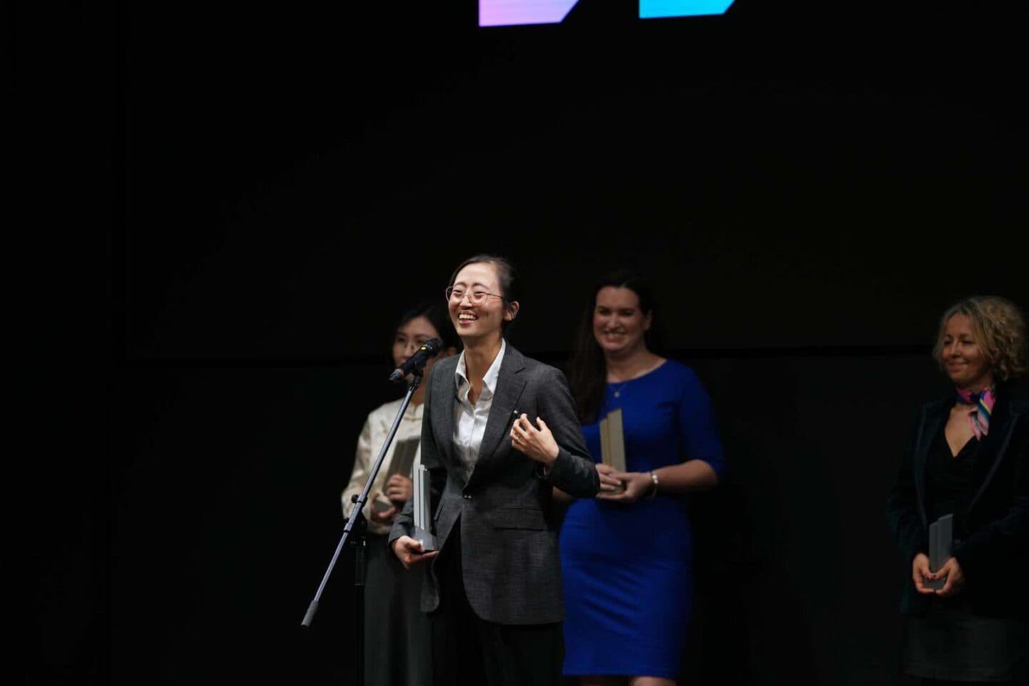
Associate Professor Jiawen Li received the Judges’ Commendation at the Sony Women in Technology Award with Nature, at the award ceremony in Tokyo last week. She is the only Australian researcher among the four winners.
“It’s really exciting … to be recognised by a big-name technology company and publisher, and be seen as innovators, not just as women innovators,” she says.
Li’s work is worth celebrating. For more than a decade, she has been developing tiny imaging devices that can probe deep inside the body in a minimally invasive way. The real-time physiological data can help medical procedures from heart treatment to IVF.
Let’s shed some light on the journey to inventing this life-saving tech.

The tech behind the genius
Since childhood, Li has been fascinated by biomedicine – with a biologist father and a medical doctor mother, it was hard to avoid developing a keen interest. Combined with a talent for maths and physics, this took her to work at the interface of engineering and medicine.
“I don’t really enjoy doing the pure engineering,” she admits. “I don’t care how fast the optical fibre transmits the signal … But if we can help patients, if we can help people live better and healthier – those are the things that really make me passionate.”
She currently leads the intravascular imaging program at the University of Adelaide’s School of Electrical and Mechanical Engineering and Institute for Photonics and Advanced Sensing. Her team works closely with clinicians, biologists, physicists, chemists and engineers to develop the technology needed to improve cardiovascular health. This requires understanding the needs of the clinicians, then translating it into engineering language and problem-solving.

For example, the team has developed a tool to allow cardiologists to peer into a patient’s blood vessels, accurately identify high-risk plaques, and thus assess their risk for a heart attack.
The imaging device is flexible and ultrathin, only as wide as a few strands of human hair, and it was made possible by marrying optical fibre technology with nanoscale 3D printing. The device functions as an endoscope: a medical tool designed to give a view of the internal parts of the body.
This endoscope can image a patient’s vascular arteries with “high sensitivity and near-cellular-resolution that is not possible with existing technologies,” Li says.
She adds that this will “answer fundamental questions at cellular level of how plaques evolve, how they cause heart attacks, and how they respond to different treatment”.
She and colleagues are working towards commercialising this device here in Australia.
Expanding the senses
Li has already developed endoscopes that function beyond just the optical, in order to gather a wide range of information about the body.
During her PhD at the University of California, Irvine, Li explains that she developed a hair-thin endoscope that “combined the optical and acoustic method together – combined our eye function (how we see) with our ear function (how we hear) to improve the diagnosis of heart disease”.

The device allows clinicians to peer within a coronary artery and receive both optical and ultrasound images in real-time. This helps determine where to place stents (small mesh tubes used to hold open narrowed passages, such as coronary arteries).
This endoscope has since been commercialised and is available in the US, Canada and China, among other countries.

“After that, I started exploring different things and combining, for example, eye and nose – so see it but also smell [to get] chemical information,” Li says.
This chemical information, such as measuring pH value, has applications in fields beyond cardiovascular health. Li suggests it could be used during the egg-collection stage of IVF.
“We can use this imaging function to look at where [the egg] is, but also use the chemical function to know whether it’s a healthy or a mature egg,” she says – and thus avoid complications.
Another sense to incorporate is touch. In 2018, Li’s team created a world-first device that was able to measure temperature and capture images at the same time, with applications in helping researchers prevent drug-induced overheating of the brain.
Game-changing 3D printing
But what sets the most recent endoscope apart is the fact that it is 3D printed.
When Li worked on the early endoscopes during her PhD, their imaging performance was limited by fabrication methods.

“Since I moved to Australia, we have been working out the 3D printing to make all those small, tiny devices to be able to have unprecedented image quality and sensitivity,” she says.
Li and team have developed 3D micro-printing technology for their applications by collaborating with a German company Nanoscribe and the University of Stuttgart. This means they don’t have to reinvent the wheel – Nanoscribe and the University of Stuttgart do a lot of the heavy lifting in developing the 3D printing techniques, Li explains, while her team is “good at looking at what exactly the medical application is, how we are translating the language for a medical doctor into real device requirements, and improving their design as well”.
These printing techniques now allow them to create high-resolution devices smaller than previously possible, and in a quick and reliable manner.
“Now with the 3D printing you can upgrade to a next level of accuracy and how clear or how sharp the images are,” Li says.
When asked about the future of her research, Li is full of both practical and blue-sky ideas. But on the near horizon, she’s excited about an upcoming paper about how cardiologists are using her endoscopes.
“Getting their feedback is the key,” she says. “At this stage, it’s not so much engineering for the purpose of engineering. It’s more for what exactly [clinicians] need, how they want it to be more deployable, how they want it easier to use, how this actually fits the clinical workflow.”

Three other international researchers were recognised at the 2025 Sony Women in Technology Award with Nature:
- Amanda Randles from Duke University in Durham, North Carolina, for developing a digital twin of coronary arteries;
- Kiana Aran from the University of California San Diego, for integrating CRISPR technology with an electronic chip, designed to rapidly detect diseases;
- And Yating Wan of King Abdullah University of Science and Technology in Thuwal, Saudi Arabia, for her work developing photonic chips for data communication and processing.
For more information on the award, visit their website.
